
Read and download the NCERT Class 11 Biology Nervous Control and Coordination Important Notes. Designed for 2026-27, this advanced study material provides Class 11 Biology students with detailed revision notes, sure-shot questions, and detailed answers. Prepared by expert teachers and they follow the latest CBSE, NCERT, and KVS guidelines to ensure you get best scores.
Advanced Study Material for Class 11 Biology Chapter 18 Neural Control and Coordination
To achieve a high score in Biology, students must go beyond standard textbooks. This Class 11 Chapter 18 Neural Control and Coordination study material includes conceptual summaries and solved practice questions to improve you understanding.
Class 11 Biology Chapter 18 Neural Control and Coordination Notes and Questions
Nervous System:-
The nervous system of all animals is composed of highly specialised cells called neurons which can detect, receive and transmit different kinds of stimuli.
HUMAN NERVOUS SYSTEM :-
The human nervous system is divided into two parts:
(i) Central Nervous System (CNS)
(ii) Peripheral Nervous System (PNS)
The CNS includes the brain and the spinal cord and is the site of information processing and control. The PNS comprises of all the nerves of the body associated with the CNS (brain and spinal cord). The nerve fibres of the PNS are of two types:
(a) Afferent Fibres
(b) Efferent Fibres
The afferent nerve fibres transmit impulses from tissues/organs to the CNS and the efferent fibres transmit regulatory impulses from the CNS to the concerned peripheral tissues/organs.
The PNS is divided into two divisions called somatic nervous system and autonomic nervous system. The somatic nervous system relays impulses from the CNS to skeletal muscles while the autonomic nervous system transmits impulses from the CNS to the involuntary organs and smooth muscles of the body. The autonomic nervous system is further classified into sympathetic nervous system and parasympathetic nervous system.
NEURON AS STRUCTURAL AND FUNCTIONAL UNIT OF NERVOUS SYSTEM
A neuron is a microscopic structure composed of three major parts, namely, cell body, dendrites and axon.
Cell Body: The cell body contains cytoplasm with typical cell organelles and certain granular bodies called Nissl’s granules.
Dendrites: Short fibres which branch repeatedly and project out of the cell body also contain Nissl’s granules and are called dendrites. These fibres transmit impulses towards the cell body.
Axon: The axon is a long fibre, the distal end of which is branched. Each branch terminates as a bulb-like structure called synaptic knob which possess synaptic vesicles containing chemicals called neurotransmitters. The axons transmit nerve impulses away from the cell body to a synapse or to a neuro-muscular junction.
Based on the number of axon and dendrites, the neurons are divided into three types:
(a) Multipolar (with one axon and two or more dendrites; found in the cerebral cortex),
(b) Bipolar (with one axon and one dendrite, found in the retina of eye) and
(c) Unipolar (cell body with one axon only; found usually in the embryonic stage).
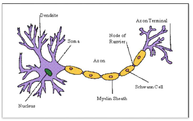
There are two types of axons, namely, myelinated and nonmyelinated. The myelinated nerve fibres are enveloped with Schwann cells, which form a myelin sheath around the axon. The gaps between two adjacent myelin sheaths are called nodes of Ranvier. Myelinated nerve fibres are found in spinal and cranial nerves. Unmyelinated nerve fibre is enclosed by a Schwann cell that does not form a myelin sheath around the axon, and is commonly found in autonomous and the somatic nervous systems.
Generation and Conduction of Nerve Impulse
Neurons are excitable cells because their membranes are in a polarized state. Different types of ion channels are present on the nervous membrane. These ion channels are selectively permeable to different ions. When a neuron is not conducting any impulse, i.e., resting, the axonal membrane is comparatively more permeable to potassium ions (K+) and nearly impermeable to sodium ions (Na+). Similarly, the membrane is impermeable to negatively charged proteins present in the axoplasm. Consequently, the axoplasm inside the axon contains high concentration of K+ and negatively charged proteins and low concentration of Na+.
In contrast, the fluid outside the axon contains a low concentration of K+, a high concentration of Na+ and thus forms a concentration gradient. These ionic gradieNeurons are excitable cells because their membranes are in a polarized state. Different types of ion channels are present on the nervous membrane. These ion channels are selectively permeable to different ions. When a neuron is not conducting any impulse, i.e., resting, the axonal membrane is comparatively more permeable to potassium ions (K+) and nearly impermeable to sodium ions (Na+). Similarly, the membrane is impermeable to negatively charged proteins present in the axoplasm. Consequently, the axoplasm inside the axon contains high concentration of K+ and negatively charged proteins and low concentration of Na+.
In contrast, the fluid outside the axon contains a low concentration of K+, a high concentration of Na+ and thus forms a concentration gradient. These ionic gradients across the resting membrane are maintained by the active transport of ions by the sodium-potassium pump which transports 3 Na+ outwards for 2 K+ into the cell. As a result, the outer surface of the axonal membrane possesses a positive charge while its inner surface becomes negatively charged and therefore is polarised. The electrical potential difference across the resting plasma membrane is called as the resting potential.
Conduction of Nerve Impulse: site is reversed, and an action potential is generated at site B. Thus, the impulse (action potential) generated at site A arrives at site BWhen a stimulus is applied at a site on the polarised membrane, the membrane at the site A becomes freely permeable to Na+. This leads to a rapid influx of Na+ followed by the reversal of the polarity at that site, i.e., the outer surface of the membrane becomes negatively charged and the inner side becomes positively charged. The polarity of the membrane at the site is thus reversed and hence depolarised. The electrical potential difference across the plasma membrane at the site A is called the action potential, which is in fact termed as a nerve impulse.
At sites immediately ahead, the axon (e.g., site B) membrane has a positive charge on the outer surface and a negative charge on its inner surface. As a result, a current flows on the inner surface from site A to site B. ptic neuron, which may or may not be separated by a gap called synaptic cleft. There are two types of synapses, namely, electrical synapses and chemical synapses. At electrical synapses, the membranes of pre- and post-synaptic neurons are in very close proximity. Electrical current can flow directly from one neuron into the other across these synapses. Transmission of an impulse across electrical synapses is very similar to impulse conduction along a single axon. Impulse transmission across an electrical synapse is always faster than that across a chemical synapse. Electrical synapses are rare in our system.
At a chemical synapse, the membranes of the pre- and post-synaptic neurons are separated by a fluid-filled space called synaptic The rise in the stimulus-induced permeability to Na+ is extremely shortlived. It is quickly followed by a rise in permeability to K+. Within a fraction of a second, K+ diffuses outside the membrane and restores the resting potential of the membrane at the site of excitation and the fibre becomes once more responsive to further stimulation.
Transmission of Impulses generate a new potential in the post-synaptic neuron. The new potential developed may be either excitatory or inhibitory.
CENTRAL NERVOUS SYSTEM
The brain is the central information processing organ of our body, and acts as the ‘command and control system’. It controls the voluntary movements, balance of the body, functioning of vital involuntary organs (e.g., lungs, heart, kidneys, etc.), thermoregulation, hunger and thirst, circadian (24-hour) rhythms of our body, activities of several endocrine glands and human behaviour. It is also the site for processing of vision, hearing, speech, memory, intelligence, emotions and thoughts.
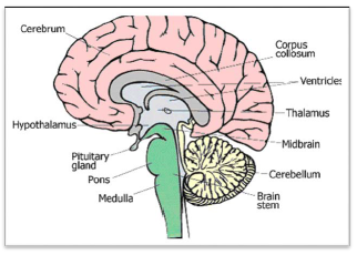
The human brain is well protected by the skull. Inside the skull, the brain is covered by cranial meninges consisting of an outer layer called dura mater, a very thin middle layer called arachnoid and an inner layer (which is in contact with the brain tissue) called pia mater. The brain can be divided into three major parts:
(i) Forebrain,
(ii) Midbrain, and
(iii) Hindbrain
Forebrain
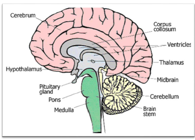
The forebrain consists of cerebrum, thalamus and hypothalamus. Cerebrum forms the major part of the human brain. A deep cleft divides the cerebrum longitudinally into two halves, which are termed as the left and right cerebral hemispheres. The hemispheres are connected by a tract of nerve fibres called corpus callosum.
The layer of cells which covers the cerebral hemisphere is called cerebral cortex and is thrown into prominent folds. The cerebral cortex is referred to as the grey matter due to its greyish appearance. The neuron cell bodies are concentrated here giving the colour.
The cerebral cortex contains motor areas, sensory areas and large regions that are neither clearly sensory nor motor in function. These regions called as the association areas are responsible for complex functions like intersensory associations, memory and communication.
Fibres of the tracts are covered with the myelin sheath, which constitute the inner part of cerebral hemisphere. They give an opaque white appearance to the layer and, hence, is called the white matter.
The cerebrum wraps around a structure called thalamus, which is a major coordinating centre for sensory and motor signaling. Another very important part of the brain called hypothalamus lies at the base of the thalamus. The hypothalamus contains a number of centres which control body temperature, urge for eating and drinking. It also contains several groups of neurosecretory cells, which secrete hormones called hypothalamic hormones.
The inner parts of cerebral hemispheres and a group of associated deep structures like amygdala, hippocampus, etc., form a complex structure called the limbic lobe or limbic system. Along with the hypothalamus, it is involved in the regulation of sexual behaviour, expression of emotional reactions (e.g., excitement, pleasure, rage and fear), and motivation.
Midbrain
The midbrain is located between the thalamus/hypothalamus of the forebrain and pons of the hindbrain. A canal called the cerebral aqueduct passess through the midbrain. The dorsal portion of the midbrain consists mainly of four round swellings (lobes) called corpora quadrigemina. Midbrain and hindbrain form the brain stem.
Hindbrain
The hindbrain comprises pons, cerebellum and medulla (also called the medulla oblongata). Pons consists of fibre tracts that interconnect different regions of the brain. Cerebellum has very convoluted surface in order to provide the additional space for many more neurons. The medulla of the brain is connected to the spinal cord. The medulla contains centres which control respiration, cardiovascular reflexes and gastric secretions.
REFLEX ACTION AND REFLEX ARC
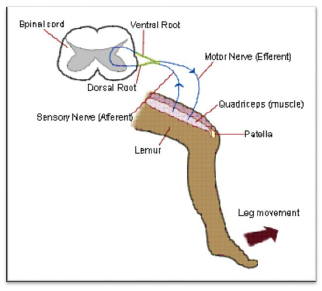
You must have experienced a sudden withdrawal of a body part which comes in contact with objects that are extremely hot, cold pointed or animals that are scary or poisonous. The entire process of response to a peripheral nervous stimulation, that occurs involuntarily, i.e., without conscious effort or thought and requires the involvment of a part of the central nervous system is called a reflex action.
The reflex pathway comprises at least one afferent neuron (receptor) and one efferent (effector or excitor) neuron appropriately arranged in a series. The afferent neuron receives signal from a sensory organ and transmits the impulse via a dorsal nerve root into the CNS (at the level of spinal cord). The efferent nueuron then carries signals from CNS to the effector. The stimulus and response thus forms a reflex arc as shown below in the knee jerk reflex.
SENSORY RECEPTION AND PROCESSING
Eye
The adult human eye ball is nearly a spherical structure. The wall of the eye ball is composed of three layers.
The external layer is composed of a dense connective tissue and is called the sclera. The anterior portion of this layer is called the cornea.
The middle layer, choroid, contains many blood vessels and looks bluish in colour. The choroid layer is thin over the posterior two-thirds of the eye ball, but it becomes thick in the anterior part to form the ciliary body.
The ciliary body itself continues forward to form a pigmented and opaque structure called the iris which is the visible coloured portion of the eye.
The eye ball contains a transparent crystalline lens which is held in place by ligaments attached to the ciliary body. In front of the lens, the aperture surrounded by the iris is called the pupil. The diameter of the pupil is regulated by the muscle fibres of iris.
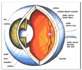
The inner layer is the retina and it contains three layers of cells – from inside to outside – ganglion cells, bipolar cells and photoreceptor cells.
There are two types of photoreceptor cells, namely, rods and cones. These cells contain the light-sensitive proteins called the photopigments. The daylight (photopic) vision and colour vision are functions of cones and the twilight (scotopic) vision is the function of the rods. The rods contain a purplish-red protein called the rhodopsin or visual purple, which contains a derivative of Vitamin A.
In the human eye, there are three types of cones which possess their own characteristic photopigments that respond to red, green and blue lights. The sensations of different colours are produced by various combinations of these cones and their photopigments. When these cones are stimulated equally, a sensation of white light is produced.
The optic nerves leave the eye and the retinal blood vessels enter it at a point medial to and slightly above the posterior pole of the eye ball. Photoreceptor cells are not present in that region and hence it is called he blind spot. At the posterior pole of the eye lateral to the blind spot, here is a yellowish pigmented spot called macula lutea with a central pit called the fovea. The fovea is a thinned-out portion of the retina where only the cones are densely packed. It is the point where the visual acuity (resolution) is the greatest.
The space between the cornea and the lens is called the aqueous chamber and contains a thin watery fluid called aqueous humor. The pace between the lens and the retina is called the vitreous chamber and is filled with a transparent gel called vitreous humor.
Mechanism of Vision
• The light rays in visible wavelength focussed on the retina through the ornea and lens generate potentials (impulses) in rods and cones.
• Light induces dissociation of the retinal from opsin resulting in changes in the structure of the opsin. This causes membrane permeability changes. As a result, potential differences are generated in he photoreceptor cells. This produces a signal that generates action potentials in the ganglion cells through the bipolar cells
• These action potentials (impulses) are transmitted by the optic nerves to the visual cortex area of the brain, where the nervous impulses are analysed and the image formed on the retina is recognised based on earlier memory and experience.
The Ear
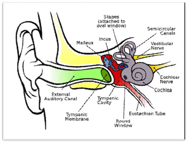
The ears perform two sensory functions, hearing and maintenance of body balance. Anatomically, the ear can be divided into three major sections called the outer ear, the middle ear and the inner ear.
Outer Ear: The outer ear consists of the pinna and external auditory meatus (canal). The pinna collects the vibrations in the air which produce sound. The external auditory meatus leads inwards and extends up to the tympanic membrane (the ear drum). There are very fine hairs and wax-secreting sebaceous glands in the skin of the pinna and the meatus. The tympanic membrane is composed of connective tissues covered with skin outside and with mucus membrane inside.
Middle Ear: The middle ear contains three ossicles called malleus, incus and stapes which are attached to one another in a chain-like fashion.
The malleus is attached to the tympanic membrane and the stapes is attached to the oval window of the cochlea. The ear ossicles increase the efficiency of transmission of sound waves to the inner ear.
An Eustachian tube connects the middle ear cavity with the pharynx. The Eustachian tube helps in equalising the pressures on either sides of the ear drum.
Inner Ear: The fluid-filled inner ear called labyrinth consists of two parts, the bony and the membranous labyrinths. The bony labyrinth is a series of channels. Inside these channels lies the membranous labyrinth, which is surrounded by a fluid called perilymph. The membranous labyrinth is filled with a fluid called endolymph. The coiled portion of the labyrinth is called cochlea.
The membranes constituting cochlea, the reissner’s and basilar, divide the surounding perilymph filled bony labyrinth into an upper scala vestibuli and a lower scala tympani. The space within cochlea called scala media is filled with endolymph. At the base of the cochlea, the scala vestibuli ends at the oval window, while the scala tympani terminates at the round window which opens to the middle ear.
The organ of corti is a structure located on the basilar membrane which contains hair cells that act as auditory receptors. The hair cells are present in rows on the internal side of the organ of corti. The basal end of the hair cell is in close contact with the afferent nerve fibres. A large number of processes called stereo cilia are projected from the apical part of each hair cell. Above the rows of the hair cells is a thin elastic membrane called tectorial membrane.
The inner ear also contains a complex system called vestibular apparatus, located above the cochlea. The vestibular apparatus is composed of three semi-circular canals and the otolith organ consisting of the saccule and utricle. Each semi-circular canal lies in a different plane at right angles to each other. The membranous canals are suspended in the perilymph of the bony canals. The base of canals is swollen and is called ampulla, which contains a projecting ridge called crista ampullaris which has hair cells. The saccule and utricle contain a projecting ridge called macula. The crista and macula are the specific receptors of the vestibular apparatus responsible for maintenance of balance of the body and posture.
NCERT Solution for Class 11 Biology Nervous Control and Coordination
Question 1. Briefly describe the structure of the following:
(a) Brain (b) Eye (c) Ear
Answer: (a) Structure of Brain
The human brain is well protected by the skull. Inside the skull, the brain is covered by cranial meninges consisting of an outer layer called dura mater, a very thin middle layer called arachnoid and an inner layer (which is in contact with the brain tissue) called pia mater. The brain can be divided into three major parts:
(i) Forebrain,
(ii) Midbrain, and
(iii) Hindbrain
Forebrain
The forebrain consists of cerebrum, thalamus and hypothalamus. Cerebrum forms the major part of the human brain. A deep cleft divides the cerebrum longitudinally into two halves, which are termed as the left and right cerebral hemispheres. The hemispheres are connected by a tract of nerve fibres called corpus callosum.
The cerebral cortex contains motor areas, sensory areas and large regions that are neither clearly sensory nor motor in function. These regions called as the association areas are responsible for complex functions like intersensory associations, memory and communication.
The cerebrum wraps around a structure called thalamus, which is a major coordinating centre for sensory and motor signaling. Another very important part of the brain called hypothalamus lies at the base of the thalamus. The hypothalamus contains a number of centres which control body temperature, urge for eating and drinking. It also contains several groups of neurosecretory cells, which secrete hormones called hypothalamic hormones.
The inner parts of cerebral hemispheres and a group of associated deep structures like amygdala, hippocampus, etc., form a complex structure called the limbic lobe or limbic system. Along with the hypothalamus, it is involved in the regulation of sexual behaviour, expression of emotional reactions (e.g., excitement, pleasure, rage and fear), and motivation.
Midbrain
The midbrain is located between the thalamus/hypothalamus of the forebrain and pons of the hindbrain. A canal called the cerebral aqueduct passess through the midbrain. The dorsal portion of the midbrain consists mainly of four round swellings (lobes) called corpora quadrigemina. Midbrain and hindbrain form the brain stem.
Hindbrain
The hindbrain comprises pons, cerebellum and medulla (also called the medulla oblongata). Pons consists of fibre tracts that interconnect different regions of the brain. Cerebellum has very convoluted surface in order to provide the additional space for many more neurons. The medulla of the brain is connected to the spinal cord. The medulla contains centres which control respiration, cardiovascular reflexes and gastric secretions.
(b) Structure of Eye
The adult human eye ball is nearly a spherical structure. The wall of the eye ball is composed of three layers.
The external layer is composed of a dense connective tissue and is called the sclera. The anterior portion of this layer is called the cornea.
The middle layer, choroid, contains many blood vessels and looks bluish in colour. The choroid layer is thin over the posterior two-thirds of the eye ball, but it becomes thick in the anterior part to form the ciliary body.
The ciliary body itself continues forward to form a pigmented and opaque structure called the iris which is the visible coloured portion of the eye.
The eye ball contains a transparent crystalline lens which is held in place by ligaments attached to the ciliary body. In front of the lens, the aperture surrounded by the iris is called the pupil. The diameter of the pupil is regulated by the muscle fibres of iris.
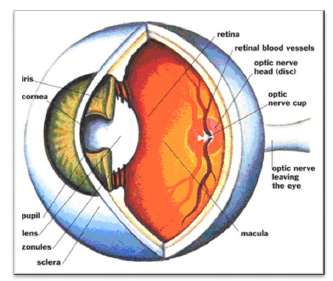
The inner layer is the retina and it contains three layers of cells – from inside to outside – ganglion cells, bipolar cells and photoreceptor cells.
There are two types of photoreceptor cells, namely, rods and cones. These cells contain the light-sensitive proteins called the photopigments. The daylight (photopic) vision and colour vision are functions of cones and the twilight (scotopic) vision is the function of the rods. The rods contain a purplish-red protein called the rhodopsin or visual purple, which contains a derivative of Vitamin A.
In the human eye, there are three types of cones which possess their own characteristic photopigments that respond to red, green and blue lights. The sensations of different colours are produced by various combinations of these cones and their photopigments. When these cones are stimulated equally, a sensation of white light is produced.
The optic nerves leave the eye and the retinal blood vessels enter it at a point medial to and slightly above the posterior pole of the eye ball. Photoreceptor cells are not present in that region and hence it is called he blind spot. At the posterior pole of the eye lateral to the blind spot, here is a yellowish pigmented spot called macula lutea with a central pit called the fovea. The fovea is a thinned-out portion of the retina where only the cones are densely packed. It is the point where the visual acuity (resolution) is the greatest.
The space between the cornea and the lens is called the aqueous chamber and contains a thin watery fluid called aqueous humor. The pace between the lens and the retina is called the vitreous chamber and is filled with a transparent gel called vitreous humor.
(c) Structure of Ear
Anatomically, the ear can be divided into three major sections called the outer ear, the middle ear and the inner ear.
Outer Ear: The outer ear consists of the pinna and external auditory meatus (canal). The pinna collects the vibrations in the air which produce sound. The external auditory meatus leads inwards and extends up to the tympanic membrane (the ear drum). There are very fine hairs and wax-secreting sebaceous glands in the skin of the pinna and the meatus. The tympanic membrane is composed of connective tissues covered with skin outside and with mucus membrane inside.
Middle Ear: The middle ear contains three ossicles called malleus, incus and stapes which are attached to one another in a chain-like fashion.
The malleus is attached to the tympanic membrane and the stapes is attached to the oval window of the cochlea. The ear ossicles increase the efficiency of transmission of sound waves to the inner ear.
An Eustachian tube connects the middle ear cavity with the pharynx. The Eustachian tube helps in equalising the pressures on either sides of the ear drum.
Inner Ear: The fluid-filled inner ear called labyrinth consists of two parts, the bony and the membranous labyrinths. The bony labyrinth is a series of channels. Inside these channels lies the membranous labyrinth, which is surrounded by a fluid called perilymph. The membranous labyrinth is filled with a fluid called endolymph. The coiled portion of the labyrinth is called cochlea.
The membranes constituting cochlea, the reissner’s and basilar, divide the surounding perilymph filled bony labyrinth into an upper scala vestibuli and a lower scala tympani. The space within cochlea called scala media is filled with endolymph. At the base of the cochlea, the scala vestibuli ends at the oval window, while the scala tympani terminates at the round window which opens to the middle ear.

The organ of corti is a structure located on the basilar membrane which contains hair cells that act as auditory receptors. The hair cells are present in rows on the internal side of the organ of corti. The basal end of the hair cell is in close contact with the afferent nerve fibres. A large number of processes called stereo cilia are projected from the apical part of each hair cell. Above the rows of the hair cells is a thin elastic membrane called tectorial membrane.
The inner ear also contains a complex system called vestibular apparatus, located above the cochlea. The vestibular apparatus is composed of three semi-circular canals and the otolith organ consisting of the saccule and utricle. Each semi-circular canal lies in a different plane at right angles to each other. The membranous canals are suspended in the perilymph of the bony canals. The base of canals is swollen and is called ampulla, which contains a projecting ridge called crista ampullaris which has hair cells. The saccule and utricle contain a projecting ridge called macula. The crista and macula are the specific receptors of the vestibular apparatus responsible for maintenance of balance of the body and posture.
Question 2. Compare the following:
(a) Central nervous system (CNS) and Peripheral nervous system (PNS)
(b) Resting potential and action potential
(c) Choroid and retina
Answer: (a) Central Nervous System and Peripheral Nervous System: The CNS includes the brain and the spinal cord and is the site of information processing and control. The PNS comprises of all the nerves of the body associated with the CNS (brain and spinal cord).
(b) Resting Potential and Action Potential: Neurons are excitable cells because their membranes are in a polarized state. Different types of ion channels are present on the nervous membrane. These ion channels are selectively permeable to different ions. When a neuron is not conducting any impulse, i.e., resting, the axonal membrane is comparatively more permeable to potassium ions (K+) and nearly impermeable to sodium ions (Na+). Similarly, the membrane is impermeable to negatively charged proteins present in the axoplasm. Consequently, the axoplasm inside the axon contains high concentration of K+ and negatively charged proteins and low concentration of Na+.
In contrast, the fluid outside the axon contains a low concentration of K+, a high concentration of Na+ and thus forms a concentration gradient. These ionic gradients across the resting membrane are maintained by the active transport of ions by the sodium-potassium pump which transports 3 Na+ outwards for 2 K+ into the cell. As a result, the outer surface of the axonal membrane possesses a positive charge while its inner surface becomes negatively charged and therefore is polarised. The electrical potential difference across the resting plasma membrane is called as the resting potential.
When a stimulus is applied at a site on the polarised membrane, the membrane at the site becomes freely permeable to Na+. This leads to a rapid influx of Na+ followed by the reversal of the polarity at that site, i.e., the outer surface of the membrane becomes negatively charged and the inner side becomes positively charged. The polarity of the membrane at the site is thus reversed and hence depolarised. The electrical potential difference across the plasma membrane at the site A is called the action potential, which is in fact termed as a nerve impulse.
(c) Choroid and Retina: The middle layer, choroid, contains many blood vessels and looks bluish in colour. The choroid layer is thin over the posterior two-thirds of the eye ball, but it becomes thick in the anterior part to form the ciliary body.
The inner layer is the retina and it contains three layers of cells – from inside to outside – ganglion cells, bipolar cells and photoreceptor cells.
Question 3. Explain the following processes:
(a) Polarisation of the membrane of a nerve fibre
(b) Depolarisation of the membrane of a nerve fibre
(c) Conduction of a nerve impulse along a nerve fibre
(d) Transmission of a nerve impulse across a chemical synapse
Answer: (a) Polarisation of the membrane of a nerve fibre:
The fluid inside the membrane contains high concentration of K+ and negatively charged proteins and low concentration of Na+.
In contrast, the fluid outside the axon contains a low concentration of K+, a high concentration of Na+ and thus forms a concentration gradient.
These ionic gradients across the resting membrane are maintained by the active transport of ions by the sodium-potassium pump which transports 3 Na+ outwards for 2 K+ into the cell. As a result, the outer surface of the axonal membrane possesses a positive charge while its inner surface becomes negatively charged and therefore is polarised.
(b) Depolarisation of the membrane of a nerve fibre:
When a stimulus is applied at a site on the polarised membrane, the membrane at the site A becomes freely permeable to Na+. This leads to a rapid influx of Na+ followed by the reversal of the polarity at that site, i.e., the outer surface of the membrane becomes negatively charged and the inner side becomes positively charged. The polarity of the membrane at the site is thus reversed and hence depolarised.
(c) Conduction of a nerve impulse along a nerve fibre:
When a stimulus is applied at a site on the polarised membrane, the membrane at the site becomes freely permeable to Na+. This leads to a rapid influx of Na+ followed by the reversal of the polarity at that site, i.e., the outer surface of the membrane becomes negatively charged and the inner side becomes positively charged.
At sites immediately ahead, the axon membrane has a positive charge on the outer surface and a negative charge on its inner surface. As a result, a current flows on the inner surface from site A to site B.
On the outer surface current flows from site B to site A to complete the circuit of current flow. Hence, the polarity at the site is reversed, and an action potential is generated at site B. Thus, the impulse (action potential) generated at site A arrives at site B.
The sequence is repeated along the length of the axon and consequently the impulse is conducted.
The rise in the stimulus-induced permeability to Na+ is extremely shortlived. It is quickly followed by a rise in permeability to K+. Within a fraction of a second, K+ diffuses outside the membrane and restores the resting potential of the membrane at the site of excitation and the fibre becomes once more responsive to further stimulation.
(d) Transmission of a nerve impulse across chemical synapse:
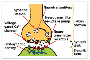
At a chemical synapse, the membranes of the pre- and post-synaptic neurons are separated by a fluid-filled space called synaptic cleft. Chemicals called neurotransmitters are involved in the transmission of impulses at these synapses. The axon terminals contain vesicles filled with these neurotransmitters. When an impulse (action potential) arrives at the axon terminal, it stimulates the movement of the synaptic vesicles towards the membrane where they fuse with the plasma membrane and release their neurotransmitters in the synaptic cleft. The released neurotransmitters bind to their specific receptors, present on the post-synaptic membrane. This binding opens ion channels allowing the entry of ions which can generate a new potential in the post-synaptic neuron. The new potential developed may be either excitatory or inhibitory.
Question 4. Give a brief account of:
(a)
Mechanism of vision
(b) Mechanism of hearing
Answer: (a) Mechanism of Vision
• The light rays in visible wavelength focussed on the retina through the ornea and lens generate potentials (impulses) in rods and cones.
• Light induces dissociation of the retinal from opsin resulting in changes in the structure of the opsin. This causes membrane permeability changes. As a result, potential differences are generated in he photoreceptor cells. This produces a signal that generates action potentials in the ganglion cells through the bipolar cells.
• These action potentials (impulses) are transmitted by the optic nerves to the visual cortex area of the brain, where the nervous impulses are analysed and the image formed on the retina is recognised based on earlier memory and experience.
(b) Mechanism of Hearing:
The outer part of the ear collects sound. That sound pressure is amplified through the middle portion of the ear and, in land animals, passed from the medium of air into a liquid medium. The change from air to liquid occurs because air surrounds the head and is contained in the ear canal and middle ear, but not in the inner ear. The inner ear is hollow, embedded in the temporal bone, the densest bone of the body. The hollow channels of the inner ear are filled with liquid, and contain a sensory epithelium that is studded with hair cells. The microscopic "hairs" of these cells are structural protein filaments that project out into the fluid. The hair cells are mechanoreceptors that release a chemical neurotransmitter when stimulated. Sound waves moving through fluid push the filaments; if the filaments bend over enough it causes the hair cells to fire. In this way sound waves are transformed into nerve impulses.
Question 5. Answer briefly:
(a) How do you perceive the colour of an object?
(b) Which part of our body helps us in maintaining the body balance?
(c) How does the eye regulate the amount of light that falls on the retina.
Answer: (a) Cones are responsible for color vision. They require brighter light to function than rods require. There are three types of cones, maximally sensitive to long-wavelength, medium-wavelength, and short-wavelength light (often referred to as red, green, and blue, respectively, though the sensitivity peaks are not actually at these colors). The color seen is the combined effect of stimuli to, and responses from, these three types of cone cells.
(b) The Inner ear has three semi-circular canals forming cochlea. Cochlea is responsible for maintaining the body balance.
(c) The pupil in the eye functions like an aperture. This dilates in case of low light and constricts in case of intense light thereby regulating the amount of light falling on the retina.
(a)
Question 6. Differentiate between:
(a) Myelinated and non-myelinated axons
(b) Dendrites and axons
(c) Rods and cones
(d) Thalamus and Hypothalamus
(e) Cerebrum and Cerebellum
Answer: (a) Myelinated and non-myelinated axons: The myelinated nerve fibres are enveloped with Schwann cells, which form a myelin sheath around the axon. The gaps between two adjacent myelin sheaths are called nodes of Ranvier. Myelinated nerve fibres are found in spinal and cranial nerves. Unmyelinated nerve fibre is enclosed by a Schwann cell that does not form a myelin sheath around the axon, and is commonly found in autonomous and the somatic nervous systems.
(b) Dendrites: Short fibres which branch repeatedly and project out of the cell body also contain Nissl’s granules and are called dendrites. These fibres transmit impulses towards the cell body.
Axon: The axon is a long fibre, the distal end of which is branched. Each branch terminates as a bulb-like structure called synaptic knob which possess synaptic vesicles containing chemicals called neurotransmitters. The axons transmit nerve impulses away from the cell body to a synapse or to a neuro-muscular junction.
(c) Rods and Cones: There are two types of photoreceptor cells, namely, rods and cones. These cells contain the light-sensitive proteins called the photopigments. The daylight (photopic) vision and colour vision are functions of cones and the twilight (scotopic) vision is the function of the rods. The rods contain a purplish-red protein called the rhodopsin or visual purple, which contains a derivative of Vitamin A.
(d) Thalamus and Hypothalamus: The cerebrum wraps around a structure called thalamus, which is a major coordinating centre for sensory and motor signaling. Another very important part of the brain called hypothalamus lies at the base of the thalamus. The hypothalamus contains a number of centres which control body temperature, urge for eating and drinking. It also contains several groups of neurosecretory cells, which secrete hormones called hypothalamic hormones.
(e) Cerebrum and Cerebellum: The cerebrum is located in the forebrain while cerebellum is located in the hind brain.
Question 7. The region of the vertebrate eye, where the optic nerve passes out of the retina, is called the
(a) fovea
(b) iris
(c) blind spot
(d) optic chaisma
Answer: (c) Blind Spot
Question 8. Distinguish between:
(a) afferent neurons and efferent neurons
(b) impulse conduction in a myelinated nerve fibre and unmyelinated nerve fibre
(c) aqueous humor and vitreous humor
(d) blind spot and yellow spot
(e) cranial nerves and spinal nerves.
Answer: (a) The afferent nerve fibres transmit impulses from tissues/organs to the CNS and the efferent fibres transmit regulatory impulses from the CNS to the concerned peripheral tissues/organs.
(b) The evolutionary need for the fast and efficient transduction of electrical signals in nervous system resulted in appearance of myelin sheaths around neuronal axons. Myelin sheath reduces membrane capacitance and increases membrane resistance in the inter-node intervals, thus allowing a fast, saltatory movement of action potentials from node to node. Myelination is found mainly in vertebrates, but an analogous system has been discovered in a few invertebrates, such as some species of shrimp. Not all neurons in vertebrates are myelinated; for example, axons of the neurons comprising autonomous (vegetative) nervous system are not myelinated in general.
The conduction velocity v of myelinated neurons varies roughly linearly with axon diameter whereas the speed of unmyelinated neurons varies roughly as the square root of diameter. Myelin has two important advantages: fast conduction speed and energy efficiency. Also, since the ionic currents are confined to the nodes of Ranvier, there is far fewer ions "leak" across the membrane, saving metabolic energy. This saving is a significant selective advantage, since the human nervous system uses approximately 20% of the body's metabolic energy.
(c) The space between the cornea and the lens is called the aqueous chamber and contains a thin watery fluid called aqueous humor. The pace between the lens and the retina is called the vitreous chamber and is filled with a transparent gel called vitreous humor.
(d) The optic nerves leave the eye and the retinal blood vessels enter it at a point medial to and slightly above the posterior pole of the eye ball. Photoreceptor cells are not present in that region and hence it is called he blind spot. At the posterior pole of the eye lateral to the blind spot, here is a yellowish pigmented spot called macula lutea with a central pit called the fovea. The fovea is a thinned-out portion of the retina where only the cones are densely packed. It is the point where the visual acuity (resolution) is the greatest.
(e) Cranial nerves are nerves that emerge directly from the brain stem in contrast to spinal nerves which emerge from segments of the spinal cord. Peripheral nerves are separated to achieve segmental innervation, cranial nerves are divided to serve one or a few specific functions in wider anatomical territories.
Free study material for Biology
CBSE Class 11 Biology Chapter 18 Neural Control and Coordination Study Material
Students can find all the important study material for Chapter 18 Neural Control and Coordination on this page. This collection includes detailed notes, Mind Maps for quick revision, and Sure Shot Questions that will come in your CBSE exams. This material has been strictly prepared on the latest 2026 syllabus for Class 11 Biology. Our expert teachers always suggest you to use these tools daily to make your learning easier and faster.
Chapter 18 Neural Control and Coordination Expert Notes & Solved Exam Questions
Our teachers have used the latest official NCERT book for Class 11 Biology to prepare these study material. We have included previous year examination questions and also step-by-step solutions to help you understand the marking scheme too. After reading the above chapter notes and solved questions also solve the practice problems and then compare your work with our NCERT solutions for Class 11 Biology.
Complete Revision for Biology
To get the best marks in your Class 11 exams you should use Biology Sample Papers along with these chapter notes. Daily practicing with our online MCQ Tests for Chapter 18 Neural Control and Coordination will also help you improve your speed and accuracy. All the study material provided on studiestoday.com is free and updated regularly to help Class 11 students stay ahead in their studies and feel confident during their school tests.
FAQs
Our advanced study package for Chapter 18 Neural Control and Coordination includes detailed concepts, diagrams, Mind Maps, and explanation of complex topics to ensure Class 11 students learn as per syllabus for 2026 exams.
The Mind Maps provided for Chapter 18 Neural Control and Coordination act as visual anchors which will help faster recall during high-pressure exams.
Yes, teachers use our Class 11 Biology resources for lesson planning as they are in simple language and have lot of solved examples.
Yes, You can download the complete, mobile-friendly PDF of the Biology Chapter 18 Neural Control and Coordination advanced resources for free.
Yes, our subject matter experts have updated the Chapter 18 Neural Control and Coordination material to align with the rationalized NCERT textbooks and have removed deleted topics and added new competency-based questions.