
Download the latest CBSE Class 12 Biology Respiration Notes in PDF format. These Class 12 Biology revision notes are carefully designed by expert teachers to align with the 2026-27 syllabus. These notes are great daily learning and last minute exam preparation and they simplify complex topics and highlight important definitions for Class 12 students.
Revision Notes for Class 12 Biology Respiration
To secure a higher rank, students should use these Class 12 Biology Respiration notes for quick learning of important concepts. These exam-oriented summaries focus on difficult topics and high-weightage sections helpful in school tests and final examinations.
Respiration Revision Notes for Class 12 Biology
(b) Respiratory region :
• Epithelial lining of maxillo turbinals and inferior turbinals / Ethmoturbinals is composed of PSCGE in which goblet cells are present, which secret mucous, and makes nasal passage
moist.It act as a air conditioning and filtering unit in breathing.
(c) Olfactory region :
• Epithelial linings of nasoturbinals called neurosensory epithelium which receive smell sensation.
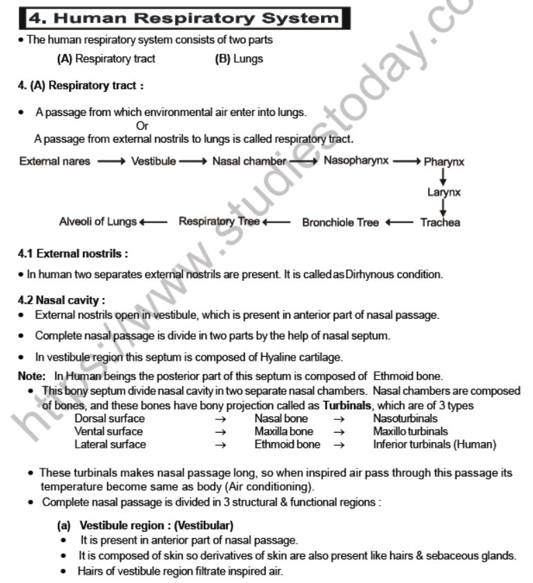
4.3 Internal nares = (choanae)
• Nasal chamber open in nasopharynx through internal nares :

4.4 Pharynx
• Due to presence of palate in mammals, anterior part of pharynx is divide in two chambers.
(1) On Dorsal surface nasopharynx
(2) On ventral surface oropharynx
• Posteriorly both these chambers open in pharynx, so pharynx is a common chamber for food and air. Therefore mammals can breath through mouth.
• In posterior part of pharynx 2 openings are present :
(1) Upper opening is called Gullet through which pharynx open in oesophagus.
(2) Lower opening is called Glottis through which pharynx open in Larynx/Trachea.
• On glottis leaf shape septum Epiglottis (thin elastic cartilage) is present which close glottis at the time of swallowing. So at this moment breathing stops or breathing rate become zero.
• Pharynx is only body part in which both food and air passage intersect each-other.
4.5 LARYNX (voice box: voice producing organ) :
• It is present in the anterior part of Trachea, so is considered as modification of Trachea.
• It is a bone like str. Which is composed of two cartilage (In structure), Four cartilage (In shape), Nine cartilage (In number)
• These cartilage of larynx attach by ligaments & membranes.

(1) Thyroid cartilage :
• Largest cartilage of Larynx.
• Composed of hyaline cartilage.
• It is C-shaped cartilage so incomplete cartilage.
• Ventrally it is broad & laterally it is narrow but absent on dorsal surface.
• So this cartilage complete almost ventral surface and little part of each lateral surface of larynx.
• On anterior part of Thyroid epiglottis is attached which is composed of elastic cartilage.
• In males of human ventral surface of Thyroid makes a process called as Adam’s apple.
(2) Cricoid cartilage :
• Composed of hyaline cartilage.
• It is signet ring shape cartilage. (so complete)
• It is present just below Thyroid cartilage.
• Dorsally it is broad and ventrally it is narrow. So this cartilage complete maximum. Dorsal surface and remaining part of each lateral surface of Larynx.
(3) Arytenoids Cartilage :
• Two in number so paired cartilage.
• Composed of hyaline cartilage.
• These are pyramid shape cartilage which are present just above cricoid cartilage in dorsal surface of Larynx.
• One end of vocal cords is attached with arytenoids and another end is attached with Thyroid cartilage.
(4) Cartilage of Santorini :
• Two in number (paired).
• It is composed of Elastic cartilage.
• They are present in the form of node like structure at the end of arytenoids. So they are considered as Bands of Arytenoids.
• When epiglottis close glottis it fall on cartilage of santorini. So these cartilage protect epiglottis.
(5) Other cartilages :
• Cuneiform and Corniculate are also present.
Vocal cords : In Larynx two pairs of vocal cords are present
(1) Anterior vocal cords : false vocal cord :
• They are composed of membranes.
• They are pink in colour.
• They do not help in Phonation.
(2) Posterior pair : True vocal cord :
• They are composed of yellow fibrous C.T., so they are yellow in colour.
• Usually they are present in relax position, so when air pass through vocal cord, no sound is produced.
• By the contraction in laryngeal muscle vocal cord comes in stretch position, so when air pass through these vocal cords, due to vibration, sound is produced in the form of Laryngeal voice (AA and E) which is converted into true speech by the help of lips and tongue.
• It is due to presence of well developed speech centre.
• This centre was started from ‘Neanderthal man’.
Note :
(i) Sound Production : Sound is produced by true vocal cords. When expired air is passed through the true vocal cords under pressure from the lungs, the vocal cords are set into vibration, which results in the production of sound.
(ii) Pitch of a sound : It is determined by the tension on the vocal cords, greater the tension - higher the pitch.
(iii) Quality of voice : It depends on the resonators above the larynx, namely the pharynx, mouth, bucca cavity and paranasal sinuses.
(iv) Quality of sound : It is controlled by the muscles of the soft palate, tongue, floor of mouth, cheeks, lips and jaws.
4.6 Trachea :
• Its 12 cm long tube with diameter 2.5 cm, which is present in complete length of neck and upto middle part of thoracic cavity (at the level of 5th thoracic vertebra divide into right and left primary bronchi).
• In complete length of Trachea 16-20 ‘C-shaped’ cartilaginous rings are present which are composed of hyaline cartilage.
• These rings are incomplete on dorsal surface of Trachea which prevent trachea to collapse.
• In the absence of cartilage on dorsal surface Trachealis muscles are present which help in dialation of Trachea during forceful breathing.
• In the histology of wall of Trachea there are four layers.
(1) Mucosa : 3 sub layers
(a) Epithelium : PSCGE
(b) Lamina propria : Reticular fibrous C.T.
(c) Muscularis mucosa : Longitudinal & circular muscle
(2) Submacosa : Aerolar C.T.
(3) Cartilagenous layer : C shape rings of hyaline cartilage
(4) Tunica Adventia : White fibrous C.T.
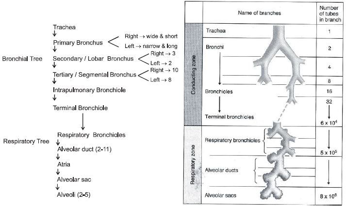
4.7 Bronchial Tree (B.T.) and Respiratory Tree (R.T.) :
• When trachea enter into thoracic cavity it divides in two branches called as primary bronchus. Branches of primary bronchus upto Terminal bronchioles make Bronchial Tree.
• Terminal bronchioles divide to form respiratory bronchiole and its branches make Respiratory Tree.
Note:
(i) Cartilagenous rings are present in the wall of bronchial tree while absent in respiratory tree.
(ii) Gaseous exchange occur in Respiratory tree while absent in Bronchial Tree.
(iii) Volume of air which is present in bronchial tree is a part of Dead space volume.

(1) Thyroid cartilage :
• Largest cartilage of Larynx.
• Composed of hyaline cartilage.
• It is C-shaped cartilage so incomplete cartilage.
• Ventrally it is broad & laterally it is narrow but absent on dorsal surface.
• So this cartilage complete almost ventral surface and little part of each lateral surface of larynx.
• On anterior part of Thyroid epiglottis is attached which is composed of elastic cartilage.
• In males of human ventral surface of Thyroid makes a process called as Adam’s apple.
(2) Cricoid cartilage :
• Composed of hyaline cartilage.
• It is signet ring shape cartilage. (so complete)
• It is present just below Thyroid cartilage.
• Dorsally it is broad and ventrally it is narrow. So this cartilage complete maximum. Dorsal surface and remaining part of each lateral surface of Larynx.
(3) Arytenoids Cartilage :
• Two in number so paired cartilage.
• Composed of hyaline cartilage.
• These are pyramid shape cartilage which are present just above cricoid cartilage in dorsal surface of Larynx.
• One end of vocal cords is attached with arytenoids and another end is attached with Thyroid cartilage.
(4) Cartilage of Santorini :
• Two in number (paired).
• It is composed of Elastic cartilage.
• They are present in the form of node like structure at the end of arytenoids. So they are considered as Bands of Arytenoids.
• When epiglottis close glottis it fall on cartilage of santorini. So these cartilage protect epiglottis.
(5) Other cartilages :
• Cuneiform and Corniculate are also present.
Vocal cords : In Larynx two pairs of vocal cords are present
(1) Anterior vocal cords : false vocal cord :
• They are composed of membranes.
• They are pink in colour.
• They do not help in Phonation.
(2) Posterior pair : True vocal cord :
• They are composed of yellow fibrous C.T., so they are yellow in colour.
• Usually they are present in relax position, so when air pass through vocal cord, no sound is produced.
• By the contraction in laryngeal muscle vocal cord comes in stretch position, so when air pass through these vocal cords, due to vibration, sound is produced in the form of Laryngeal voice (AA and E) which is converted into true speech by the help of lips and tongue.
• It is due to presence of well developed speech centre.
• This centre was started from ‘Neanderthal man’.
Note :
(i) Sound Production : Sound is produced by true vocal cords. When expired air is passed through the true vocal cords under pressure from the lungs, the vocal cords are set into vibration, which results in the production of sound.
(ii) Pitch of a sound : It is determined by the tension on the vocal cords, greater the tension - higher the pitch.
(iii) Quality of voice : It depends on the resonators above the larynx, namely the pharynx, mouth, bucca cavity and paranasal sinuses.
(iv) Quality of sound : It is controlled by the muscles of the soft palate, tongue, floor of mouth, cheeks, lips and jaws.
4.6 Trachea :
• Its 12 cm long tube with diameter 2.5 cm, which is present in complete length of neck and upto middle part of thoracic cavity (at the level of 5th thoracic vertebra divide into right and left primary bronchi).
• In complete length of Trachea 16-20 ‘C-shaped’ cartilaginous rings are present which are composed of hyaline cartilage.
• These rings are incomplete on dorsal surface of Trachea which prevent trachea to collapse.
• In the absence of cartilage on dorsal surface Trachealis muscles are present which help in dialation of
Trachea during forceful breathing.
• In the histology of wall of Trachea there are four layers.
(1) Mucosa : 3 sub layers
(a) Epithelium : PSCGE
(b) Lamina propria : Reticular fibrous C.T.
(c) Muscularis mucosa : Longitudinal & circular muscle
(2) Submacosa : Aerolar C.T.
(3) Cartilagenous layer : C shape rings of hyaline cartilage
(4) Tunica Adventia : White fibrous C.T.
4.7 Bronchial Tree (B.T.) and Respiratory Tree (R.T.) :
• When trachea enter into thoracic cavity it divides in two branches called as primary bronchus. Branches of primary bronchus upto Terminal bronchioles make Bronchial Tree.
• Terminal bronchioles divide to form respiratory bronchiole and its branches make Respiratory Tree.
Note:
(i) Cartilagenous rings are present in the wall of bronchial tree while absent in respiratory tree.
(ii) Gaseous exchange occur in Respiratory tree while absent in Bronchial Tree.
(iii) Volume of air which is present in bronchial tree is a part of Dead space volume.
Note :
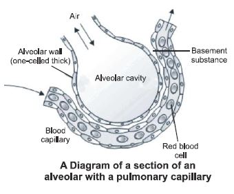
(i) Alveoli is structural and functional unit of lungs.
(ii) In two lungs 300 million alveoli are present.
(iii) Wall of alveoli consist of two layers:
(1) Outer layer is composed of sheet of yellow fibrous C.T. in which network of blood capillaries is present.
(2) Inner layer is composed of simple squamous epithelium. Squamous cells of alveoli are called as Pneumocytes.
(iv) Most of these pneumocytes help in gaseous exchange while few pneumocytes which are larger in size they secret Lecithin which is also called surfactant which prevent alveolar collapse. The lecithin lining
(1) lowers the surface tension of alveoli and keeps them open preventing collapse.
(2) Speeds up the diffusion of gases between air and blood.
(3) kills bacteria that may reach the lungs.
4 (B) Lungs :
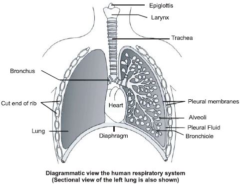
• In human two light, spongy & pink in colour lungs are present.
• Around lungs pleural cavity is present which is a space between parietal pleura & visceral pleura .
• Both pleura are composed of simple squamous epithelium.
• In this cavity pleural fluid is filled which is 2 ml in amount.
• By infection when amount of this fluid is increase it is called as pleurisy which causes chest pain & breathing become painful.
• Painful or difficult breathing condition called dyspnoea.
• Space between two lungs is called as Mediastinum in which heart, oesophagus, Aorta, posterior venacava, Thymus gland & lymph vessels are present.
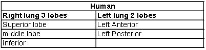
• Right lung is bigger than left lung.
• In inner surface of left lung a permanent concavity is present called as Cardiac notch, which is formed by compression of heart
Note:
(i) Thoracic cage :
Covering of thoracic cavity makes thoracic cage.
Anterior surface : Neck & Clavicle
Posterior surface : Diaphragm
Ventral surface : Sternum & Ribs
Dorsal surface : Vertebral column & Ribs
Lateral surface : Ribs
(ii) Diaphragm :
• A muscular septum which is found in only mammals.
exception : crocodiles
• It is dome-shape septum which divide body cavity in two parts.
(1) Upper thoracic cavity
(2) Lower abdominal cavity.
• In centre of diaphragm a central Tendon is present which is pierced by 3 structures :
(1) Oesophagus
(2) Aorta
(3) Posterior vana cava
• In diaphragm radial muscles are present which originate from periphery and inserted in centre of diaphragm.
• By the contraction in these muscles, diaphragm become flattened in shape, so volume of thoracic cavity increase, therefore diaphragm help in only inspiration.
(iii) Intercostals Muscles (ICM) :
• Space between two ribs is called as intercostals space in which two types of muscles are present.
(1) External Intercostals Muscles (EICM) :
• The fibres of an EICM originate from Posterior border of the dorsal part of a rib and insert upon anterior border of the ventral part of the rib behind crossing the IICM at right angle.
Contraction of these muscles assist in inspiration.
(2) Internal intercostals muscles [IICM] :
• They originates from dorsal surface of lower rib and inserted on ventral surface of upper rib.
• By the contraction in these muscles ribs and sternum shifts downwards and inwards.They help in only forceful expiration. So they are under control of cerebrum.
Mechanism of Respiration
Breathing :
• In take of fresh air from environment and expulsion of fawl air from lungs is called breathing, so there are two steps in breathing :
(a) Inspiration
(b) Expiration
(a) Inspiration :
• In take of fresh air
• It is an active process.
• Completes in 2 sec.
• Inspiration occur by contraction in inspiratory muscles
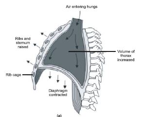
which are of two types :
(i) Radial muscles of diaphragm
(ii) EICM
• When radial muscles contract diaphragm become flattened in shaped so volume of thoracic cavity increase between anterior & posterior surface.
• When EICM contract sternum comes outward and ribs goes upward.
• By the contraction in both muscles volume of thoracic cavity increase and intrapulmonary pressure decrease by 1-3 mm Hg. So, environmental air enter into lungs through respiratory tract called inspiration
(b) Expiration :
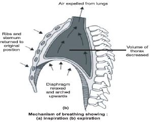
• Explusion is flow of air.
• It is a passive process.
• It takes 3 sec.
• Expiration occur by relaxation in inspiratory muscles.
• By relaxation in radial muscle diaphragm become normal dome-shape.
• When EICM relax, then ribs and sternum comes to their normal position.
So by relaxation in both muscles volume of thoracic cavity decrease and intrapulmonary pressure increase 1-3 mm Hg. So expulsion of air occur through respiratory tract called expiration.
Note : During exercise forceful expiration take place in which with relaxation of inspiratory muscles, contraction of expiratory or respiratory muscles also takes place.
• These muscles are of two types :
(1) Abdominal muscles
(2) IICM
• When abdominal muscles contract, visceral organs of abdominal cavity push diaphragm upwards. So, it become more dome shape.
• When IICM contract ribs and sternum shift downward and inward.
• By contraction in both muscles volume of thoracic cavity decrease more and intrapulmonary pressure increase more, so more expiration occur through respiratory tract called forceful expiration.
Note :
(i) Normal breathing [Eupnoea] is called as Abdominal breathing which is under control of medulla.
Role of Diaphragm - 75 %
ICM - 25%
(ii) Forceful breathing is called as Thoracic breathing which is under control of cerebrum.
(iii) A pregnant female usually breaths by Thoracic breathing
Role of Diaphragm - 25 %
ICM - 75%
Spirometry : (Pulmonary air Volumes & Capacities)
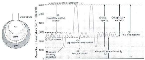
(1) Tidal Volume (TV) :
Volume of air which is inspired or expired in normal breathing. It is 500 ml.
(2) Inspiratory reserve volume [IRV] :
Volume of air which inspired forceful beyond Tidal volume. It is 3000 ml
(3) Expiratory reserve volume [ERV] :
Volume of air which expired forcefully beyond Tidal volume. It is 1100 ml
(4) Residual volume [RV] :
Volume of air which always remain in lungs after forceful expiration. It can not expired in any condition.
It is 1200 ml. So, Functional residual volume is ERV + RV = 2300 ml
(5) Vital capacity of lungs [VC] :
Volume of air which expired forcefully after forceful inspiration.
Vital capacity = IRV + ERV + TV
= 3000 + 1100 + 500
= 4600 ml
(6) Inspiratory Capacity [IC] : IRV + TV
= 3000 + 500 = 3500 ml
(7) Functional Residual Capacity (FRC) :
Volume of air that will remain in the lungs after a normal expiration. This includes ERV + RV
1100 + 1200 = 2300 ml
(8) Total capacity of lungs :
Volume of air which can be filled in lungs.
Total capacity = Vital capacity + RV
= 4600 + 1200 = 5800 ml
(9) Dead Space Volume :
Complete volume of fresh air do not take part in gaseous exchange, while a part of this air retain in respiratory tract from external nostrils to terminal bronchiole called dead space volume. It is 150ml.
(10) Minute respiratory volume :
Volume of air which is inspired or expired per minute in normal breathing.
It is 500 x 12 = 6000 ml.
(11) Alveolar ventilation :
Volume of fresh air which take part in gaseous exchange per minute.
It is 350 x 12 = 4200 ml.
(12) Expiratory Capacity (EC) :
Total volume of air a person can expire after a normal inspiration. This includes tidal volume and expiratory
reserve volume (TV + ERV)
Exchange of gases
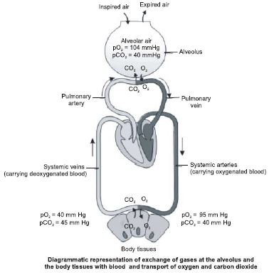
Gaseous exchange between lungs and blood and blood with tissue fluid occur by simple diffusion due to partial pressure difference.
Gaseous Exchange in Lungs
At alveoli level gaseous exchange occur through respiratory membrane which is 0.2 mm thick and consist of three layers :
(1) Alveolar epithelium
(2) Basement membrane
(3) Capillary endothelium
• The respiratory membrane has a limit of gaseous exchange between alveoli and pulmonary blood. It is called diffusing capacity. The diffusing capacity is defined as the volume of gas, that diffuses through the membrane per minute for a pressure difference of 1 mm Hg.
• It is further dependent on the solubility of the diffusing gases.
• As the solubility of CO2 is 20-25 times higher than that of O2, the amount CO2 that can diffuse through the diffusion membrane per unit difference in partial pressure is much higher compared to that of O2
• The partial pressure of oxygen (PO2) in the alveoli is higher (104 mm Hg) than that in the deoxygenated blood in the capillaries of the pulmonary arteries (40 mm Hg). As the gases diffuse from a higher to a lower concentration, the movement of oxygen is from the alveoli to the blood.
• The reverse is the case in relation to carbon dioxide.
The partial pressure of carbon dioxide (PCO2) is higher in deoxygenated blood (45 mm Hg) than in alveoli (40 mm Hg). Therefore, carbon dioxide passes from the blood to the alveoli.Partial Preseures (In mm Hg) of O2 and CO2 at Different Parts involved in Diffusion
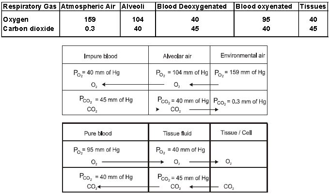
7.2 Gaseous Exchange in Tissues :
• The partial pressure of oxygen in the oxygenated blood is higher (95mm Hg) than that of the tissue cells (40mm Hg) and the partial pressure of carbondioxide in the oxygenated blood is lesser (40mm Hg) than that of the tissue cells (45mm Hg).
• Therefore, oxygen diffuses from the capillary blood to the tissue cells through tissue fluid and carbon dioxide diffuses form the tissue cells of the capillary blood through tissue fluid.
Transport of gases
8.1 Transport of oxygen :
(a) 97% to 99% oxygen is transported as oxyhaemoglobin and
(b) 1-3 % O2 transported as dissolve form in water of plasma.
• Haemoglobin is a red coloured pigment present in RBC. O2 can bind with Hb in a reversible manner to form oxyhaemoglobin.
• One molecule of Hb contains four molecule of O2.
Note:
(i) OXYHAEMOGLOBIN DISSOCIATION CURVE :
• This curve is plotted between partial pressure of oxygen and percentage saturation of haemoglobin with O2.
• It is sigmoid in shape.
• P50 : It is defined as partial pressure of O2 at which Hb is half saturated.
(1) In oxygenated blood haemoglobin is 97% saturated with oxygen in which partial pressure of oxygen is 100 mm Hg.
(2) Deoxygenated blood in which partial pressure of O2 is 40 mm Hg, haemoglobin is 75% saturated with oxygen. So, in a single circulation Hb release 22% [20-25%] oxygen to tissue fluid.
OR
100 ml blood release 5 ml oxygen to tissue fluid.
(3) During vigorous exercise, due to more demand of oxygen haemoglobin delivers 75% oxygen to tissue fluid
OR
100 ml blood release 15 ml to tissue fluid.
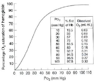
• If body temperature increase, CO2 conc. increase, pH decrease, Acidity increase.Then dissocia tion curve of oxyhaemoglobin shift to right means more dissociation of oxyhaemoglobin take place.
• Oxygen dissociation curve of myoglobin is hyperbolic. Function of myoglobin is to store oxygen in muscles
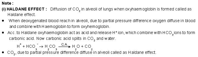
(ii) BOHR EFFECT :
In the active tissues, there is increased concentration of CO2, which also increases H+ ion (CO2 react with water to form carbonic acid). Both these factors induce oxyhaemoglobin to give up more oxygen. This phenomenon is called Bohr effect or Bohr shift (Bohr, 1904). Bohr effect plays crucial role in enhancing the release of oxygen in the tissues.
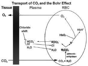
Transport of CO2 : Occur in three forms :
(a) As dissolve form : 7% CO2 is transported as dissolved form in water of plasma.
(b) As carbamino haemoglobin : 23% Hb. NH2+ CO2 → Hb. NH. COOH
This percentage of CO2 combine with amino group of globin protein of haemoglobin to form carbaminohaemoglobin
(c) As bicarbonate compound (70% CO2)
• This percentage of CO2 diffuse in RBC and dissolve in water of cytoplasm to form carbonic acid.Formation of carbonic acid is occur in presence of carbonic anhydrase enzyme which increase rate of formation of carbonic acid 5000 times.
• Carbonic acid now dissociates into H+ ions and Bicarbonate (HCO3–) ions.
• To regulate pH of blood haemoglobin act as buffer and combine with H+ ion to form Haemoglobinicacid.
• Bicarbonate ions are highly diffusible ions. They diffuse in plasma from RBC and combine with Na+ions to form Sodium Bicarbonate.
• To maintain ion equilibrium, in the same of HCO3
– ions Cl– ions also diffuse in RBC from plasma. It is called chloride shift or Hamburger phenomena.
• Cl– ions combine with K+ ion to form potassium chloride.
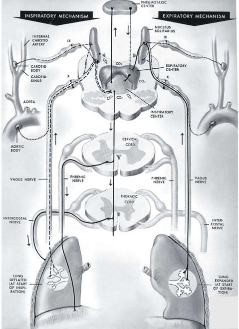
Regulation of Respiration
Nervous control of breathing :
• There are four centers in brain which regulate breathing :
(a) Inspiratory centre
(b) Apneustic center
(c) Pneumotaxic centre
(d) Expiratory centre
(a) Inspiratory centre :
It is located on dorsal surface of medulla, so called as Dorsal group of neuron.
• This centre is under chemical control.
• When CO2 concentration increase means O2 conc. decrease, this centre stimulate and generate impulse of contraction.These impulse goes to diaphragm through phrenic nerve and through inter coastal nerve they goes to EICM. So by contraction in these muscles inspiration takes place.
(b) Apneustic center : Located in pons. Apneusis refers to inspiratory gaps. It facilitates inspiration, for example it can prolong inspiration when our O2 requirement is increased, as during exercise. In this sense the apneustic centre control depth of respiration.
(c) Pneumotaxic centre :
• It is located on dorsal surface of Pons veroli. This area control other two centers and cuts of inspiration at a certain point to make sure that inspiration does not continue too long.
• In this sense pneumotaxic center helps to control rate of respiration. For example it shuts off the hyperstimulated inspiratory centre in response to strenuous exercise preventing over inflation of lungs.
(d) Expiratory centre :
It is located on ventral surface of Medulla, so called as ventral group of neuron.
• This centre is responsible for forceful breathing.
• In this centre few group of neuron generate impulse for forceful inspiration and few group of neuron for forceful Expiration.
Note: This centre do not play any role in normal breathing
Key Concepts
Herring – Breuer Reflex :
It is a reflex which prevent overstretching or bursting of Alveoli.
• In the wall of alveoli and small bronchioles modified sensory cells are present called as stretch receptors.
• When alveoli completely filled with air, these receptors stimulate and generate impulse which reach in brain through vagus nerve and causes inhibition of inspiratory centre. So inspiration stop and expiration start.
Chemical control :
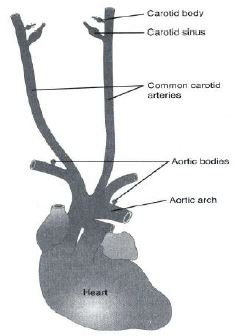
• A large number of chemoreceptors are located in carotid bodies present bilaterally in the bifurcations of the common carotid arteries. Their afferentnerve fibres pass through glassopharyngeal cranial nerves (IX th) and hence to the dorsal respiratory group of neurons in the medulla oblongata.
• Sufficient number of chemoreceptors are also located in the aortic bodies present along the arch of the arota.
Their afferent nerve fibres pass through vagus cranial nerves (Xth), and hence to the dorsal respiratory group of neurons.
• A chemosensitive area is situated adjacent to the respiratory rhythm centre which is highly sensitive to CO2 and hydrogen ions (H+ ions). Increase in these substances can activate this centre, which is turn can signal the rhythm centre to make necessary adjustments in the respiratory process by which these substances can be eliminated.
• Receptors associated with aortic arch and carotid artery also can recognise changes in CO2 and H+ ion concentration and send necessary signals to the respiratory rhythm centre for remedial actions.
• The role of oxygen in the regulation of respiratory rhythm is quite insignificant.
Disorder of Respiratory System
(1) Bronchial Asthma :
It is an allergy caused by some allergens like pollen grain, dust particle. allergen stimulate mast cells to produce histamine that causes contraction of smooth muscles of bronchi.
Symptoms : coughing, difficulty in breathing mainaly during expiration, breathing with wheezing sound, excess mucus secretion from respiratory tract that may clog bronchi and bronchiole.
(2) Emphysema :
Emphysema = inflation = full of air.
• Emphysema is an abnormal distension of bronchi, bronchiole and alveolar sac of the lungs mainaly due to cigarette smoking.
• the septa between alveoli are dissolved and elastic tissue replaced by fibrous connective tissue so that lung become non-elastic. Respiratory surface of lung reduced, bronchiole become non- elastic, alveoli remain filled with air even after exhalation.
(3) Bronchitis :
Inflammation of the bronchi due to cigaratte somking air pollutant like CO and Microbial infection
(4) Pneumonia :
It is an acute infection and inflammation of the lung alveoli by S.pneumonae, Mycoplasma, some false yeast. Infant, young ones, HIV patient are more sensitive for pneumonia.
Symptoms : The alveoli become acutely inflammated, most of the air space filled with mucus and fluid with W.B.C.
(5) Occupational lung diseases :
These disorders are caused due to exposure of potentially harmful substances, such as gas, fumes or dusts, present in the environment where a person works.
(a) Silicosis and asbestosis : These are common examples, which occur due to chronic exposure of silica and asbestos dust in the mining industry.
(b) Pneumoconiosis : It is found in coal workers.
(c) Byssinosis : It is found in workers of cotton industry.
Key Concepts
• Diptheria : Inflamation and enlarging of mucus lining of trachea, Nasopharynx, larynx, bronchi due to infection of Cornebacterium diphtheriae.
• Common cold (coryza) : A group of rhinovirus causes 40% common cold.
• Influenza : Caused by influenza virus
• Tuberculosis (TB) : Caused by Mycobacterium tuberculosis.
• Rhinitis : Inflammation of nasal mucosa.
• Pleurisy : Inflammation of Pleure (Covering of lung)
• SARS (Severe Acute Respiratory Syndrome) :
• Caused by HCV (Human carona virus) that is mutant form of influenza virus.
• First infected person reported in china on Feb. 26, 2008
• First infected person in india was prasheel verde of Goa.
Symptoms – Cold, dry cough, headache, loss of appetite, fever, hypoxia, muscular stiffness.
Diagnosis – By ELISA and rapid molecular genetic test
Mode of transmission – HCV spread through contact, respiratory secretion, cockroach.
• Carbon-Monoxide Poisoning : CO has 250 time more affinity with Hb than O2. CO combines with haemoglobin far more readily than O2, forming a relatively stable compound carboxyhaemoglobin.
This causes low supply of O2 to the body cells. It is characterised by headache, dizziness, nausea, paralysis and even death.
• Coughing : Preceded by a long-drawn and deep inspiration that is followed by a complete closure of the glottis-resulting a forcible expiration suddenly pushes glottis open and sends a blast of air through the upper respiratory passages.
• Sneezing : Spasmodic contraction of muscles of expiration forcefully expels air through the nose and mouth.
• Yawning : A prolonged inspiration through widely opened mouth producing an exaggerated depression of the lower jaw.
• Hiccough : Spasmodic contraction of the diaphragm followed by a spasmodic closure of the glottis produces a sharp inspiratory sound.
• Snoring : It is a rough rattling inspiratory noise produced by vibration of uvula or sometimes of vocal cords during sleep.
• Hay fever : An acute inflammation of the mucous membrane of upper respiratory passage.
• Asphyxia : Asphyxia is the condition characterised by combination of hypoxia and hypercapnea.
• Mountain Sickness : As the barometric pressure falls progressively with the rise in altitude, the pO2 falls proportionately in the atmospheric air. This lower the alveolar pO2 and consequenctly reduces the diffusion of oxygen from the alveolar air to the blood. As a result, oxygenation of blood is decreased progressively with the rise in altitude.
Symptoms : It includes breathlessness, headache, dizziness, irritability, nausea, vomitting, mental fatigue and a bluish tinge on the skin, nails and lips. This is called mountain sickness.
• Meaning of some terms:
Apnea – Absence of breathing
Eupnea – Normal breathing
Dyspnea – Painful breathing
Orthopnea – Inability to breathe in a horizontal position
Acapnoea – Absence of CO2 in blood
Hypocapnea – Deficiency of CO2 in blood
Hypercapnea – Excess of CO2 in blood
Hypoxaemia – Lack of O2 in arterial blood
Anoxia – Absence of O2 in tissues
Hypoxia – Lack of O2 in tissues
Tachypnea / hyperopnea – Rapid breathing
Bradypenoea / hypopnea – Slow breathing
Costal breathing – Shallow (Chest) breathing
• Breathing rate :
No. of breathings per minute.
Human - Adult → 15-18
New born → 40-50
5 years → 25
50 years → 18-25
Rabbit → 36-38
• Relation between Pulmonary ventilation and alveolar ventilation: Pulmonary ventilation is always more than alveolar ventilation.
• Blood buffers : Hb also act on buffer by taking protons and donating protons.
• Respiratory quotient (or RQ or respiratory coefficient) : It is a dimensionless number used in calculations of basal metabolic rate (BMR) when estimated from carbon dioxide production. Such measurements, like measurements of oxygen uptake, are forms of indirect calorimetry . It is measured using Ganong’s Respirometer .
The respiratory quotient (RQ) is calculated from the ratio:
RQ = CO2 eliminated /O2 consumed where the term “eliminated” refers to carbon dioxide (CO2) removed (“eliminated”) from the body.
The range of respiratory coefficients for organisms in metabolic balance usually ranges from 1.0 (representing the value expected for pure carbohydrate oxidation) to ~0.7 (the value expected for pure fat oxidation).
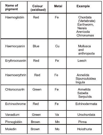
• Respiratory pigment Hemocyanin : It is a respiratory proteins in the form of metalloproteins containing two copper atoms that reversibly bind a single oxygen molecule (O2). Oxygenation causes a colour change between the colourless Cu(I) deoxygenated form and the blue Cu(II) oxygenated form. Hemocyanins carry oxygen in the hemolymph of most molluscs , and some arthropods , including the horseshoe crab , Limulus polyphemus. Most hemocyanins bind with oxygen non-cooperatively and are roughly onefourth as efficient as hemoglobin at transporting oxygen per amount of blood.
• Some Respiratory pigments with there examples:
Regulation of Respiration
Free study material for Biology
CBSE Class 12 Biology Respiration Notes
Students can use these Revision Notes for Respiration to quickly understand all the main concepts. This study material has been prepared as per the latest CBSE syllabus for Class 12. Our teachers always suggest that Class 12 students read these notes regularly as they are focused on the most important topics that usually appear in school tests and final exams.
NCERT Based Respiration Summary
Our expert team has used the official NCERT book for Class 12 Biology to design these notes. These are the notes that definitely you for your current academic year. After reading the chapter summary, you should also refer to our NCERT solutions for Class 12. Always compare your understanding with our teacher prepared answers as they will help you build a very strong base in Biology.
Respiration Complete Revision and Practice
To prepare very well for y our exams, students should also solve the MCQ questions and practice worksheets provided on this page. These extra solved questions will help you to check if you have understood all the concepts of Respiration. All study material on studiestoday.com is free and updated according to the latest Biology exam patterns. Using these revision notes daily will help you feel more confident and get better marks in your exams.
FAQs
You can download the teacher prepared revision notes for CBSE Class 12 Biology Respiration Notes from StudiesToday.com. These notes are designed as per 2026-27 academic session to help Class 12 students get the best study material for Biology.
Yes, our CBSE Class 12 Biology Respiration Notes include 50% competency-based questions with focus on core logic, keyword definitions, and the practical application of Biology principles which is important for getting more marks in 2026 CBSE exams.
Yes, our CBSE Class 12 Biology Respiration Notes provide a detailed, topic wise breakdown of the chapter. Fundamental definitions, complex numerical formulas and all topics of CBSE syllabus in Class 12 is covered.
These notes for Biology are organized into bullet points and easy-to-read charts. By using CBSE Class 12 Biology Respiration Notes, Class 12 students fast revise formulas, key definitions before the exams.
No, all study resources on StudiesToday, including CBSE Class 12 Biology Respiration Notes, are available for immediate free download. Class 12 Biology study material is available in PDF and can be downloaded on mobile.